Liver Cancer
Home / Service / Liver Cancer
Liver Cancer
Liver Cancer
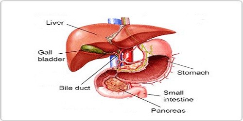
Primary liver cancer, or hepatocellular carcinoma, is the most common type of cancer originating in the liver itself. (Most tumours in the liver do not originate there; they start elsewhere in the body and spread, or metastasize, to the liver.) In the United States, primary liver cancer is relatively rare — it accounts for less than one per cent of all cancers. But worldwide, hepatocellular carcinoma is the most common solid organ tumour. This is believed to be due to widespread viral hepatitis infection, a known risk factor for primary liver cancer.
Most primary liver cancers originate in the liver’s parenchymal cells — the cells that perform most of the organ’s blood-filtering functions. Other rarer forms of primary liver cancer include peripheral cholangiocarcinoma (tumors in the sections of the bile ducts that are within the liver), sarcomas and angiosarcomas (cancer in the connective tissue of the liver), hemangioendotheliomas (tumors that arise in the blood vessels of the liver), and hepatoblastomas (a highly curable form of liver cancer most often found in children).
Hepatocellular carcinoma most commonly occurs in people whose livers have been damaged. This damage may be caused by alcohol abuse, by chronic infection with the hepatitis B or hepatitis C virus, from food contaminants called aflatoxins (though this is rare in the United States), or from metabolic diseases. Cancer can spread from the liver to other areas in the body through the blood or the lymph system, most often to the lungs, bones, and abdomen.
Risk Factors
Viral hepatitis is often a silent disease. The hepatitis virus can be present in the body for years and cause no pain or symptoms. As many as four million Americans may carry the hepatitis C virus, and most may not be aware that they are infected. Viral hepatitis is contracted through contact with infected blood or body fluids. In many cases, people became infected through blood transfusions administered before 1992 (before blood was first routinely screened for the disease). A small number of cases are still associated with recent blood transfusions. Intravenous drug users may become infected through contact with unsterilized needles. These infections are considered so serious that the U.S. Centers for Disease Control and Prevention issued guidelines in October 1998 requiring hospitals to track down and notify anyone who may have received infected blood prior to 1992.
Early in the infection hepatitis B can be treated with a combination of the anti-viral drugs alpha-interferon and ribavirin. In some cases the virus can be eradicated from the bloodstream and eliminated from the body. For this reason, doctors recommend that people at a high risk for developing the disease be screened. If the infection progresses, it can lead to chronic liver disease, or cirrhosis, a progressive disease of the liver, and, eventually, liver cancer. There is also a vaccine for hepatitis B. Doctors recommend that children, and those at high risk for developing the disease be vaccinated.
What Are The Symptoms And Signs Of Liver Cancer?
Many patients with primary liver cancer have no symptoms. In some instances, jaundice, malaise, or a general feeling of poor health, loss of appetite, weight loss, fever, fatigue, bloating, itching, swelling of the legs, or weakness may be present. Abdominal pain or discomfort may also occur.
Diagnosis
Diagnosis of primary liver cancer is generally made using blood tests, diagnostic imaging, surgical biopsy or laparoscopy, or a combination of the above. The alpha-fetoprotein blood test and ultrasound imaging of the liver are also used to screen high-risk populations (including those with hepatitis B and hepatitis C infections) for the disease. Since the risk of liver cancer is relatively low for healthy individuals, these tests are not used to screen the general population.
In some cases, diagnosis is performed invasively, by removing a small amount of tissue for a biopsy, or by laparoscopy (insertion of a small tube with an attached camera into the abdomen to survey the cancer site). Laparoscopy can also be used to remove a sample of tissue for biopsy.
Noninvasive Diagnostic Imaging Techniques
- CT (computed tomography) scanning – is useful for determining the extent of tumour growth within the gallbladder or bile duct. It can also be used to tell whether tumour cells have spread into the lymph nodes or other nearby parts of the body.
- MRI (magnetic resonance imaging) — can be used to determine if a tumour can be surgically removed. It shows the extent of tumour growth within the gallbladder or bile duct and reveals whether the tumour has invaded any blood vessels
- Magnetic resonance cholangiopancreatography (MRCP) — gives a detailed examination of the bile ducts. It is useful for determining the stage of a tumour in the bile duct.
- Ultrasound — useful for detecting the location and number of tumours as well as tumour involvement with blood vessels (tumours situated close to blood vessels may be more difficult to remove). It can also be used to distinguish a cancerous mass from a benign tumour.
Invasive Diagnostic Techniques
- Biopsy – a small amount of tissue is removed from a specific area of the body so it can be examined more closely.
- Endoscopy – the interior lining of a body cavity, such as the oesophagus, stomach, bile duct, or colon, is examined using a device called an endoscope
- Laparoscopy – allows for the examination of the abdominal cavity and its contents. A tube with an attached camera (called a laparoscope) is passed through an incision made in the abdominal wall.
- Cholangiography – a needle is inserted into the bile ducts within the liver. The ducts are injected with dye so they can be seen more clearly.
Treatment
For treatment purposes, primary liver tumours are classified in four ways. Localized and resectable tumours are found in one place and can be removed. Localized and unresectable tumours are found in one area but cannot be totally removed safely. In advanced cases, cancer has spread throughout the liver and/or to other parts of the body. In recurrent cases, cancer has returned to the liver or another part of the body after initial treatment
Ablative Therapies
Ablative therapies can be used alone or in combination with surgical removal of a tumour. For example, a patient with hepatocellular cancer who is not a candidate for surgery may first be treated with embolization to shrink the tumour so that it is small enough to make another form of ablative therapy or surgery possible.
In cryosurgery, a needle is introduced into the middle of a tumour to freeze it. Residual tumour cells can be left behind, making this method less effective than surgery. It can also be difficult to keep the tumour at temperatures low enough to completely freeze it since tumours are often near large blood vessels. Nevertheless, cryosurgery can be a very effective way to control liver tumours.
Radiation Therapy
Radiation therapy is used in selected cases to help control tumours. Radiation oncologists here use new techniques to focus the radiation beam on the tumour and spare the normal liver from injury.
Embolization
Embolization is a procedure that cuts off the blood supply to the tumour. Physicians pack a branch of the hepatic artery – the main artery that carries blood to the liver – with tiny plastic particles, cutting off most of the blood flow and depriving the tumour of life-giving oxygen.